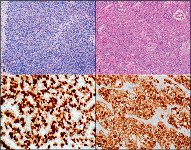
Figure 5.
Morphologic mimickers of pancreatic neuroendocrine neoplasms. A and B, Acinar cell carcinoma: relatively uniform cells forming acinar architecture with granular, eosinophilic cytoplasm. B, Trypsin immunohistochemical stain is strongly positive. C and D, Solid pseudopapillary neoplasm: sheets of polygonal cells with degenerative changes. D, The tumor cells are diffusely positive for nuclear β-catenin (hematoxylin-eosin, original magnification ×100 [A and C]; original magnification ×400 [B and D]).

