
Figure 1 |.
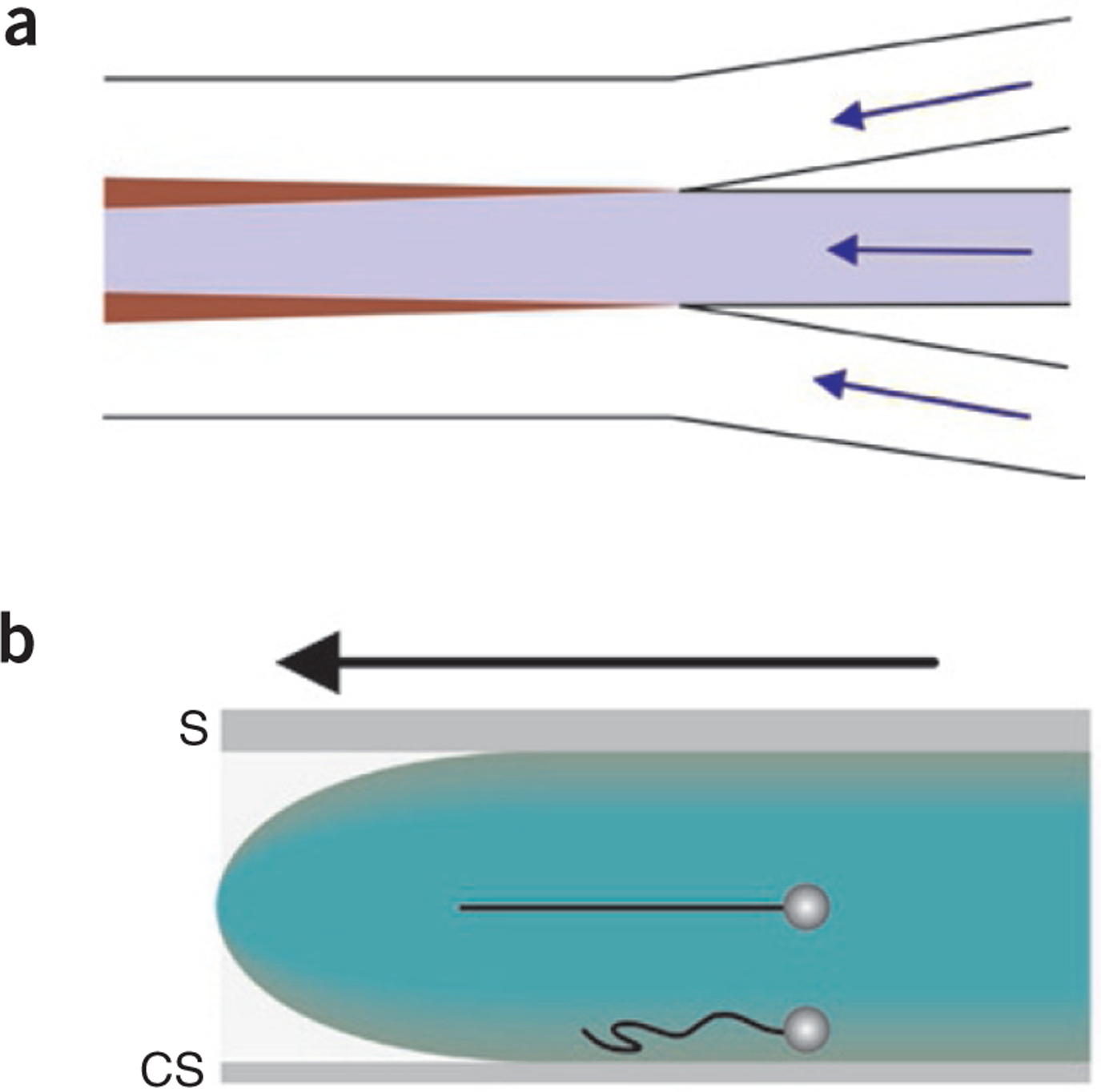
Fluid flow in multistream laminar flow cells. (a) Interchannel diffusion is the main source of mixing between adjacent fluid streams. The flow paths are indicated by blue arrows, and the individual streams are colored white, blue and white. The widening regions of transverse diffusion between channels are indicated in maroon. (b) The flow profile (green) in laminar flow cells is parabolic. The flow cell is viewed from the side with the direction of flow indicated by the black arrow. The fastest fluid velocities occur at the center of the flow cell, and the slowest occur next to the flow cell surfaces. The upper optically trapped DNA-bead complex is positioned ~18 μm from the surface, resulting in full extension of the DNA molecule (black string). Closer to the surface, DNA molecules are not fully stretched (lower DNA molecule). S, slide; CS, coverslip.