
Figure 4 |.
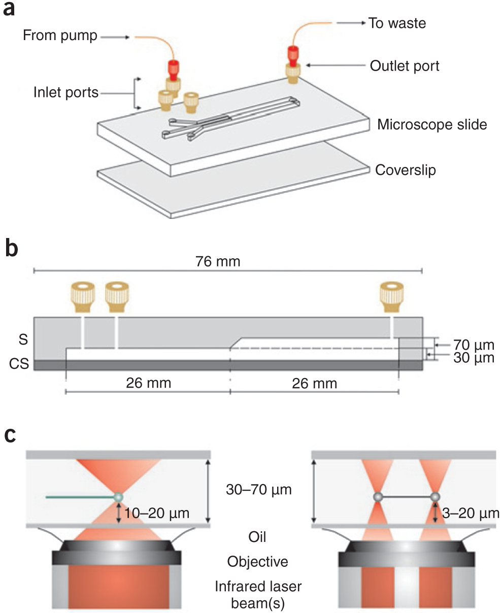
Multistream laminar flow cell for single-molecule studies. (a) A three-stream laminar flow cell constructed from glass8. The designis for an inverted microscope. Inlet channel widths are 250 μm and the common channel width is 750 μm. Machined connectors (tan; Upchurch Scientific) are attached to the slide at the holes. These are then connected to Fingertight connectors (red; Upchurch Scientific) to facilitate the attachment of PEEK tubing. Identical methods may be used to construct two-channel flow cells1,19. (A fully assembled flow cell positioned on a microscope stage is shown in Supplementary Fig. 1a.) (b) A side view of the flow cell in a showing relevant dimensions. (c) A side view of the flow cell demonstrating the trapping height from the coverslip using either one (left) or two (right) optical traps. Optical trapping and DNA visualization are carried out in the deeper portion of the flow cell in our groups.