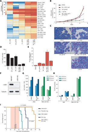
Fig. 1. LSD1 inhibition sensitizes AML cells to physiological concentrations of RA.

(A) Heat map representing the results of cell proliferation assays performed on 21 AML cell lines treated with MC_2580 (2 μM) and/or RA as indicated. Values are normalized on dimethyl sulfoxide (DMSO) treatment. The AML French-American-British classifications of each cell line are in parentheses. (B) Growth curves of NB4 APL cells treated as indicated. (C) Morphological analysis by May-Grünwald-Giemsa staining of NB4 cells treated for 96 hours in liquid culture as indicated. (D) Colony-forming ability, scored after 7 days, of 1000 NB4 cells plated in methylcellulose medium and treated with MC_2580 (2 μM) and/or 0.01 μM RA and 1 μM RA. Means and SDs of three independent experiments are shown. C.F.U., colony-forming units. (E) Analysis of CD11b mRNA levels in NB4 cells treated for 96 hours in liquid culture as indicated. Values are normalized against GAPDH (glyceraldehyde phosphate dehydrogenase) and referred to DMSO. The graph represents the mean and SD of three independent experiments. FC, fold change. (F) Western blot showing depletion of LSD1 in NB4 cells knocked out for LSD1 (LSD1 KO). WT, wild type. (G) Proliferation assays of NB4 and NB4 LSD1 KO cells after 24, 48, and 72 hours of the indicated treatments (DMSO as control). (H) Expression of CD11b by fluorescence-activated cell sorting (FACS) in NB4 and NB4 LSD1 KO cells after 24, 48, and 72 hours of the indicated treatments (DMSO as control). (I) Kaplan-Meier survival plots for mice transplanted with murine APL cells and treated as indicated (n = 6 for each treatment group). Pink shaded area indicates the duration of RA treatment (21 days, pellet), while LSD1i (DDP_38003) was administrated twice a week orally (OS) for the entire duration of RA treatment (total, six times). P values were obtained using analysis of variance (ANOVA).
