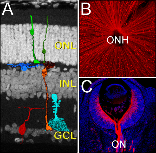
Figure 1.

Cytoarchitecture of the mammalian retina. A) The retina is organized in three main layers, the outer nuclear layer (ONL), the inner nuclear layer (INL) and the ganglion cell layer (GCL), and contains six main neuronal cell types, the cone and rod photoreceptors (light and dark green, respectively), the horizontal cells (blue), the bipolar cells (orange), the amacrine cells (light blue), and the retinal ganglion cells (red). B) All RGCs (red) send their axons to the center of the eye, a region called the optic nerve head (ONH). The image shows the RGCs of an adult flat-mounted retina. C) As axons leave the eye, they bundle to form the optic nerve (ON). The image shows a section of a E13.5 mouse retina immunolabeled with Tuj1 (red) and co-stained with DAPI (blue).
