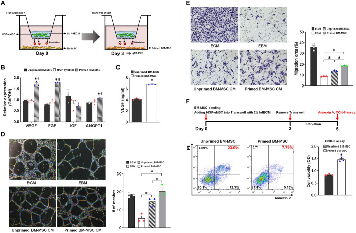
Fig. 1. In vitro characterization of primed BM-MSCs by HGF-eMSC coculture.
(A) Schematic diagram showing the in vitro coculture model of BM-MSCs with HGF-eMSCs. (B) qRT-PCR analysis of relative mRNA expression of multiple angiogenic factors in the unprimed BM-MSCs, primed BM-MSCs with HGF-eMSCs at 3 days after cocultures, or BM-MSCs receiving HGF cytokine (50 ng/ml) for 3 days. The y axis represents relative mRNA expression of target genes to glyceraldehyde-3-phosphate dehydrogenase (GAPDH). †P < 0.05 compared to unprimed BM-MSC group; n = 3 per group. (C) Measurement of secreted VEGF from unprimed or primed BM-MSCs by using ELISA. P < 0.05 compared to unprimed BM-MSC group. (D) Matrigel plug assay. The 10% hMSC-CM was treated to human umbilical cord endothelial cells (HUVECs) on Matrigel to examine the vasculogenic potential. Representative images of tubes formed on Matrigel and quantification summary. n = 3 per group. (E) Endothelial cell (EC) migration assay. The HUVECs were incubated with 10% CM harvested from hMSC cultures (hMSC-CM) or control media to evaluate the migration capability. Representative images under an inverted microscope and quantification summary are shown. *P < 0.05 compared to each group; n = 5 per group. (F) Treatment with CM from primed BM-MSCs increased cell survival after serum starvation for 5 days determined by the annexin V and cholecystokinin-8 (CCK-8) assay. *P < 0.05 compared to unprimed CM treated group; n = 3 per group. PI, propidium iodide; OD, optical density.

