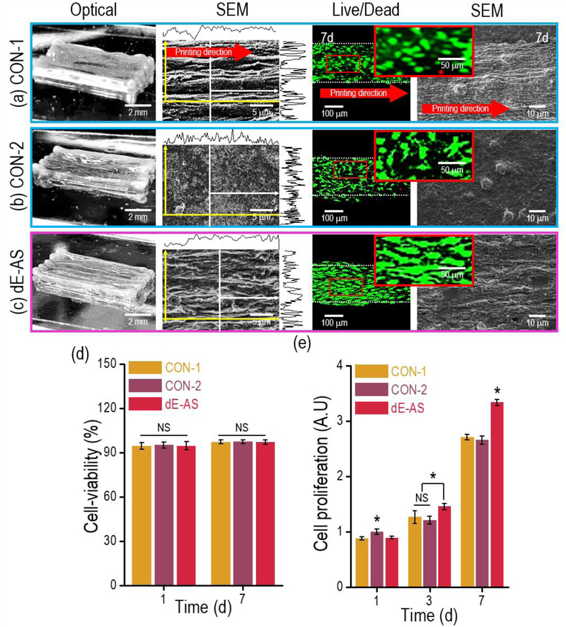
Figure 7.
Optical, SEM, and live (green)/dead (red) images for the (a) CON-1, (b) CON-2, and (c) dE-AS structures (8 × 2 × 1 mm3). (d) Cell viability for the C2C12 cells in the printed structures (CON-1, CON-2, and dE-AS) calculated using the live/dead images at 7 d (n = 9, *P < 0.05). (e) Cell proliferation of the cells in the structures, measured using an AlamarBlue staining assay (n = 9, *P < 0.05).

