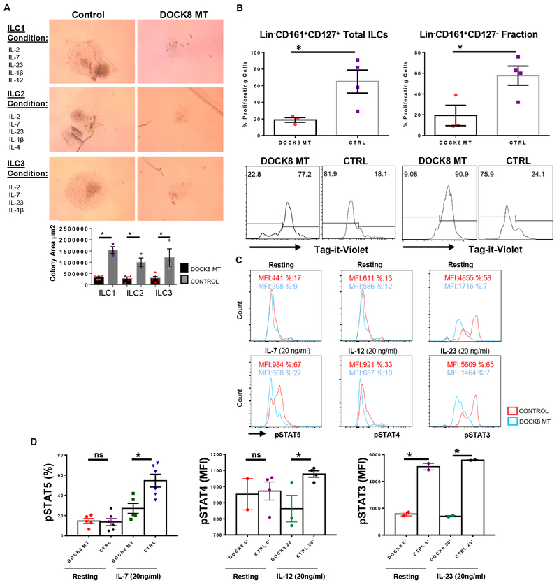
Figure 3.
Impaired proliferation and cytokine signaling of DOCK8-deficient human ILCs. A) Equal number of sorted total ILCs (CD3−Lin−CD161+CD127+) from DOCK8-deficient patients and healthy controls were cultured for 21 days with indicated cytokine ILC subset polarizing cocktails, proliferation of ILCs were quantified by area of growth. (n= 3-5 per group). B) Sorted total ILCs (CD3−Lin−CD161+CD127+) or CD3−Lin−CD161+CD127− cells were labeled with Tag-it-violet and cultured with same ILC3 subset polarizing cytokine cocktail for 5 days. * indicates p-value <0.05. DOCK8-deficient patients (n=3), healthy controls (n=4). C) IL-7R, IL-12R and IL-23R signaling in DOCK8-deficient human ILCs are impaired. Sorted ILCs (CD3−Lin−CD161+CD127+) from control and DOCK8 patients were cultured overnight and stimulated with indicated cytokines for 20 minutes. Respective STAT phosphorylation was examined via phospho-flow. Percent or MFI indicates mean fluorescence intensity. Representative histograms belong to one patient. D) Percentages of pSTAT5 in sorted total ILCs of five (5) DOCK8 MT patients and six (6) controls upon IL-7 stimulation for 20 min (left); STAT4 phosphorylation mean fluorescent intensity (MFI) upon IL-12 stimulation for 20 min (middle), total ILCs from 2 different patients used (2 technical replicates for one patient); MFI of STAT3 phosphorylation upon IL-23 stimulation for 20 min (right), (technical replicates of one patient, the other patient’s histogram was presented in Figure S3 in the Online Repository). * indicates p-value <0.05, ns: not significant.

