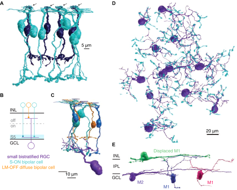
Figure 1. Serial EM reconstruction of ipRGCs and the S-cone connectome in primate retina.
(A) Four representative S-cones (arrows) and their S-ON bipolar cell (blue) and OFF midget bipolar cell (purple) contacts. (B) Small bistratified RGC circuit used for verification of S-cone and S-ON bipolar cell identification. (C) 3D reconstruction of the small bistratified RGC circuit in 1B. (D) 60 S-ON bipolar cell terminals contacting 12 small bistratified RGCs. (E) 3D reconstructions of the major ipRGC subtypes in primate retina. Note the ipRGC dendrites are monostratified but appear curved as the volume slopes away from the fovea.

