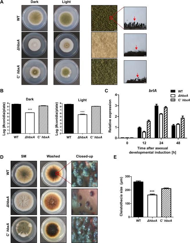
Figure 3.
Fungal development of the ΔhbxA mutant. (A) Colony morphology of WT (TNJ36), ΔhbxA (TYE14), and C’hbxA (TYE27) strains grown on MMG for 5 days at 37 °C in the light or dark. The middle panels show the magnified views of the middles of the plates (bar = 0.25 µm). The panels on the right show the morphologies of the WT and mutant conidiophores under a microscope (bar = 0.25 µm). Arrows indicate conidiophores. (B) Quantitative analysis of the conidia shown in (A). (Differences between the WT and mutants, ***p < 0.001). (C) qRT-PCR analysis for brlA mRNA levels in WT (TNJ36), ΔhbxA (TYE14), and C’hbxA (TYE27) strains after inducing asexual development. β-Actin was used as the endogenous control. (D) WT (TNJ36), ΔhbxA (TYE14), and C’hbxA (TYE27) strains were point-inoculated, and the plates were grown on SM for 7 days at 37 °C in the dark. The middle panels show the plates, which were washed with ethanol to observe the sexual structure. The panels on the right show the magnified views of the sexual structures in WT (TNJ36), ΔhbxA (TYE14), and C’hbxA (TYE27) strains (bar = 0.25 µm). (E) The sizes of the cleistothecia in WT (TNJ36), ΔhbxA (TYE14), and C’hbxA (TYE27) strains (differences between the WT and mutants, ***p < 0.001).

