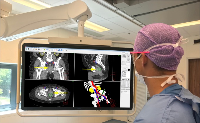
Fig. 1. Surgical navigation user interface during surgery showing the planning CT with segmentations (top-left corner: coronal view; top-right corner: sagittal view; lower-left corner: axial view) and 3D model (lower-right corner).
Visible segmentations: bones (white), arteries (red), veins (blue), ureters/kidneys (yellow), and tumor (green). The visible slice of the CT scans is based on the location of the tip of the surgical pointer which is highlighted with yellow arrows.

