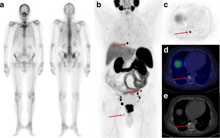
Fig. 1.
Example of a patient (PSA 44 ng/mL, Gleason score 9, T3) classified as M0 according to initial BS as shown in anterior (a) and posterior projection (b). The patient was referred for 68Ga-PSMA-11 PET/CT due to high-risk prostate cancer. The maximum intensity projection (MIP) of the 68Ga-PSMA-11 PET (c) revealed several lesions with avid 68Ga-PSMA-11 uptake, including three bone metastases marked with arrows (Th8, left iliac bone and right pubic bone). The axial 68Ga-PSMA-11 PET image of the lesion in Th8 is shown in c with a fused 68Ga-PSMA-11 PET/CT image shown in d and only a slight sclerotic change in the axial CT image (e). BVC confirmed M1 status

