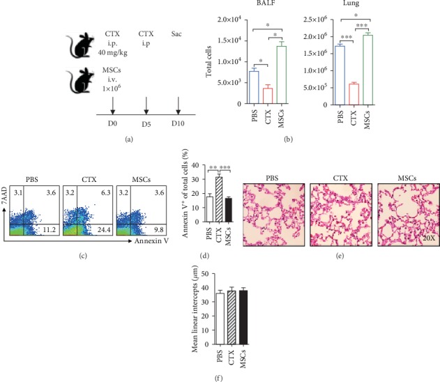
Figure 1.

CTX reduced cells in the lung, whereas MSCs increased them. (a) B6 mice were treated with MSCs or CTX and sacrificed at indicated time. i.p.: intraperitoneal; i.v.: intravenous. (b) Cells from the BALF and lungs were isolated and counted. (c, d) Apoptotic status of lung cells was determined by FACS. (e) Lung pathology was examined by H&E staining. (f) Quantitative analyses of the pulmonary alveolar sizes, as measured by MLI. Data were expressed as the means ± SEM. n = 3 mice for each treatment group; ∗p < 0.05; ∗∗p < 0.01; ∗∗∗p < 0.001. This experiment is representative of three individual experiments.
