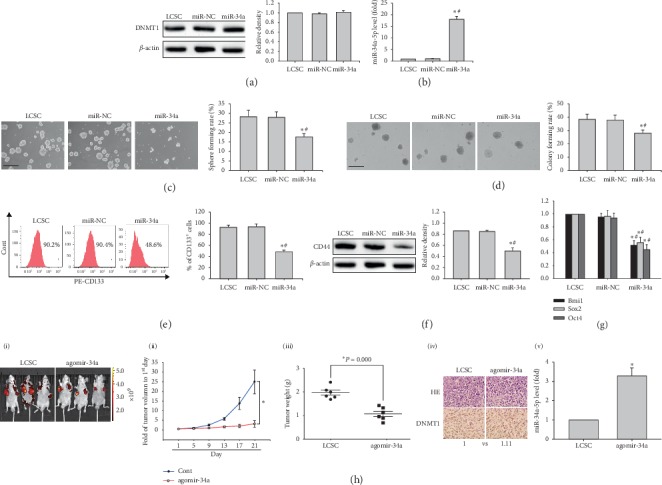
Figure 4.

Effects of miR-34a mimic on stem-like features of MHCC97H derived LCSCs. (a) DNMT1 protein amounts in LCSCs transfected with miR-34a mimic. (b) miR-34a-5p levels in LCSCs transfected with miR-34a mimic. (c, d) Representative images of spheres and colonies in LCSCs transfected with miR-34a mimic (left) (scale bar, 200 μm); sphere formation efficiencies and colony formation rates were determined (right). (e) CD133 expression in LCSCs transfected with miR-34a mimic. (f) CD44 protein amounts in LCSCs transfected with miR-34a mimic. (g) Bmi1, Sox2, and Oct4 mRNA amounts in LCSCs transfected with miR-34a mimic. (h) Images of subcutaneous xenografts from LCSCs (1 × 10⁵) expressing red fluorescent protein (RFP) treated with agomir-NC or agomir-34a (i); comparison of tumor growth curves and weights of tumors from LCSCs expressing RFP treated with agomir-NC or agomir-34a (ii, iii). ∗P < 0.05 vs. transfected with agomir-NC (n = 6). (iv) Micrographs of H&E staining (×200) and immunohistochemistry (×400) obtained under an optical microscope. (v) miR-34a-5p levels in xenografts from LCSCs transfected with agomir-34a or agomir-NC. ∗P < 0.05 vs. LCSCs (n = 6); #P < 0.05 vs. LCSCs transfected with agomir-NC (n = 6).
