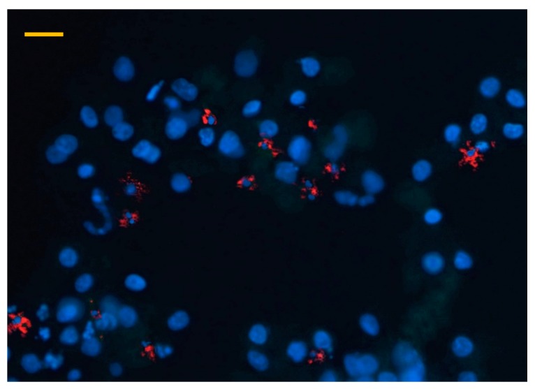
Figure 3.
Immunofluorescent detection of macrophages in a hepatocyte—nonparenchymal cell co-culture after 48 h culturing with a phycoerythrin (PE) coupled chicken macrophage specific antibody (40× magnification, bar = 40 µm). Blue colour indicates cell nuclei with DAPI staining, while red colour refers to macrophages detected with the PE conjugated antibody.

