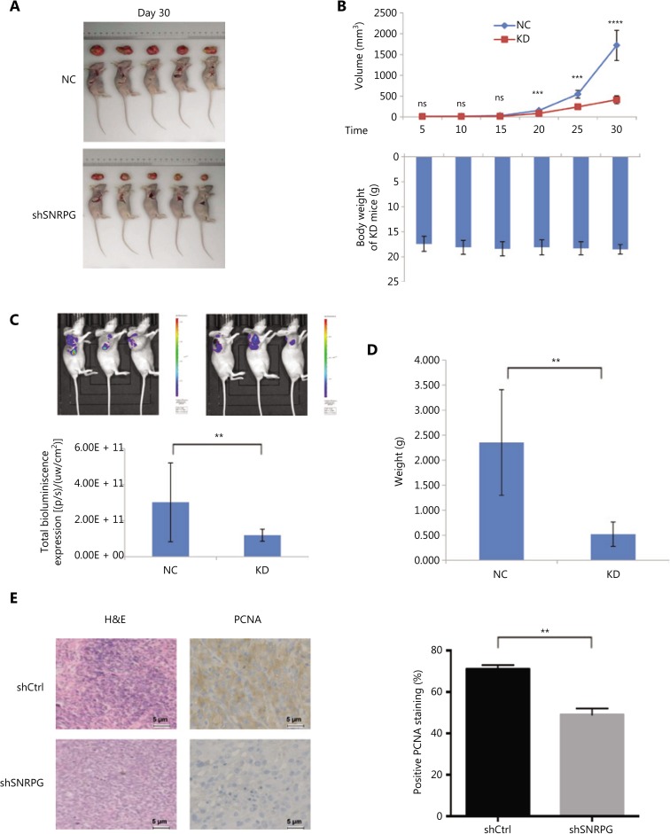
Figure 3.
Effects of SNRPG knockdown on xenograft tumorigenicity in vivo in nude mice. U87MG cells were injected into the flanks of the nude mice. (A) Tumor growth was measured on the indicated days. (B) Tumor volumes (upper panel) and body weights (lower panel) were measured on the indicated days (mean ± SD, n = 10, ns, not significant; ***P < 0.001; ****P < 0.0001 vs. the shCtrl group on the indicated days). (C) Representative bioluminescence images of shCtrl- and shSNRPG-infected U87MG cells injected into the flanks of nude mice. Mice were imaged 28 days after implantation (n = 10 in the shCtrl group and n = 20 in the shSNRPG group). In vivo imaging detection clearly showed that mice in the shCtrl groups had significantly better tumor growth delay and survival rates than mice in the shSNRPG groups. (D) Tumor weights were measured 30 days after injection (mean ± SD, n = 10, ***P < 0.001). (E) hematoxylin and eosin staining and immunohistochemistry analysis of proliferating cell nuclear antigen expression in tumors developed from U87MG/shSNRPG cells and from U87MG/shCtrl-transfected cells. Semiquantitative analysis of the stained sections was performed using light microscopy to calculate the immunoreactive score (mean ± SD, n = 3, **P < 0.001).

