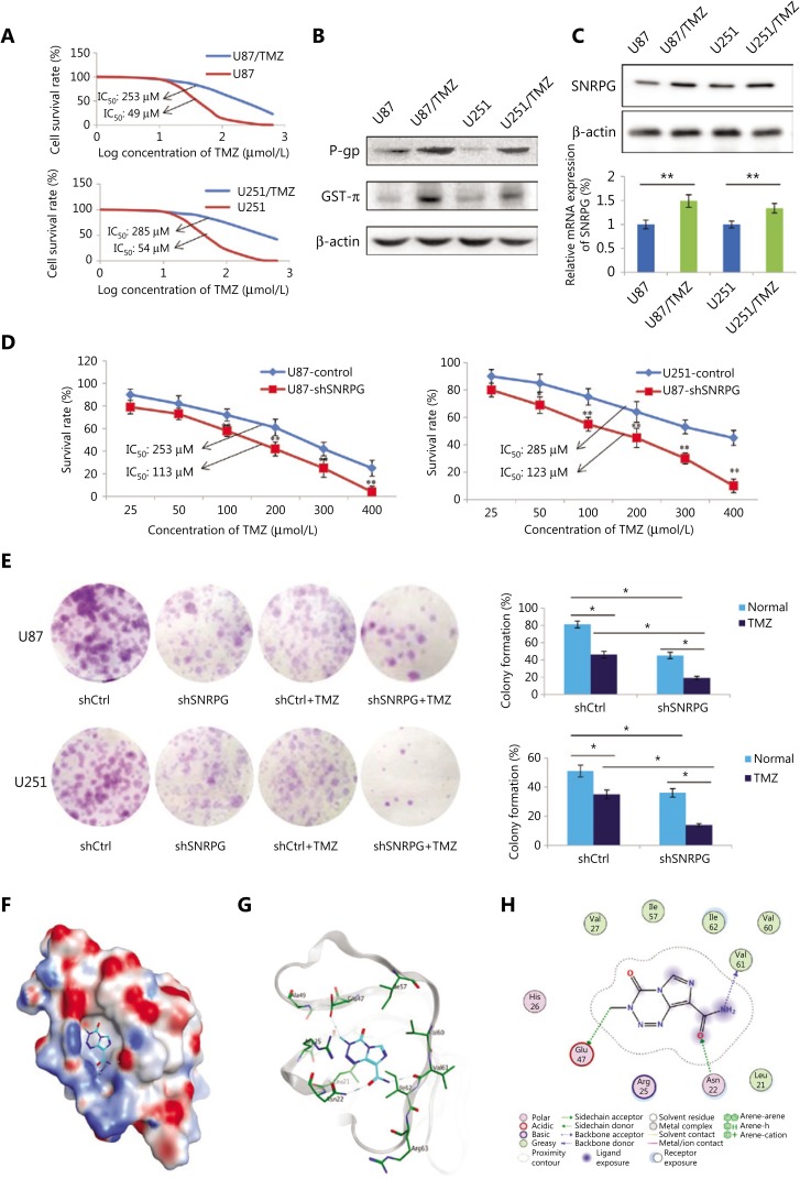
Figure 4.
SNRPG suppression sensitizes human glioblastoma multiforme cells to temozolomide (TMZ). (A) The MTT assay was used to determine the viability of U87/TMZ and U251/TMZ cells (IC50 for U87, 49 μM vs. 253 μM; IC50 for U251, 54 μM vs. 285 μM). (B) Western blot analysis of the drug resistance-related genes P-gp and GST-π in U251/TMZ and U87/TMZ cells. (C) Western blot analysis (upper panel) and RT-qPCR analysis (lower panel) of SNRPG in U251/TMZ and U87/TMZ cells. (D) The MTT assay was used to determine the cell viability after SNRPG knockdown. Downregulation of SNRPG significantly reduced the IC50 compared with that of the control group (113 μM vs. 253 μM in U87/TMZ cells; 123 μM vs. 285 μM in U251/TMZ cells; *P < 0.05; **P < 0.01). (E) A colony formation assay was used to determine the cell growth after SNRPG knockdown and TMZ treatment. *P < 0.05; **P < 0.01. The colonies were counted after staining with 0.1% crystal violet (Original magnification, 1×). (F–H) The binding mode of TMZ bound to SNRPG by molecular docking. (F) The electrostatic map surface of the SNRPG protein. Positively charged areas are colored blue, and negatively charged areas are colored red. (G) The three-dimensional binding mode of TMZ bound to SNRPG. Gray ribbons: receptor backbone. Cyan sticks: ligand TMZ. Green sticks: binding pocket residues. Blue dashes: electrostatic interactions (e.g., hydrogen bonds). (H) The two-dimensional binding mode of TMZ bound to SNRPG.

