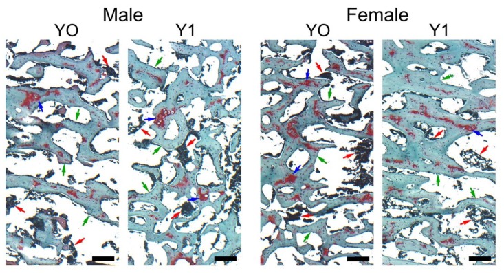
Figure 1.
Representative images of Safranine O staining carried out on formaldehyde-fixed sections of trabecular bone in the proximal section of the tibia from representative 6-week old control (Y0) and yeast-fed (Y1) Japanese quail. Green arrows indicate trabeculae, red arrows indicate bone marrow, blue arrows indicate areas of reduced mineralization of newly formed trabeculae. All the scale bars represent 100 µm.

