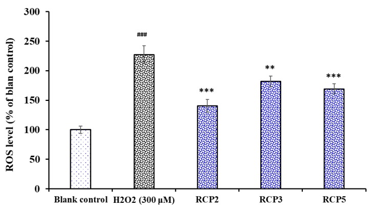
Figure 10.
Effects of RCP2, RCP3, and RCP5 on the levels of ROS in H2O2-damaged HepG2 cells at the concentration of 100 μM. The data are presented as the mean ± SD (n = 3). ### p < 0.001 versus the blank control group; *** p < 0.001 and ** p < 0.01 versus the model group (treated with H2O2 at the concentration of 300 μM).

