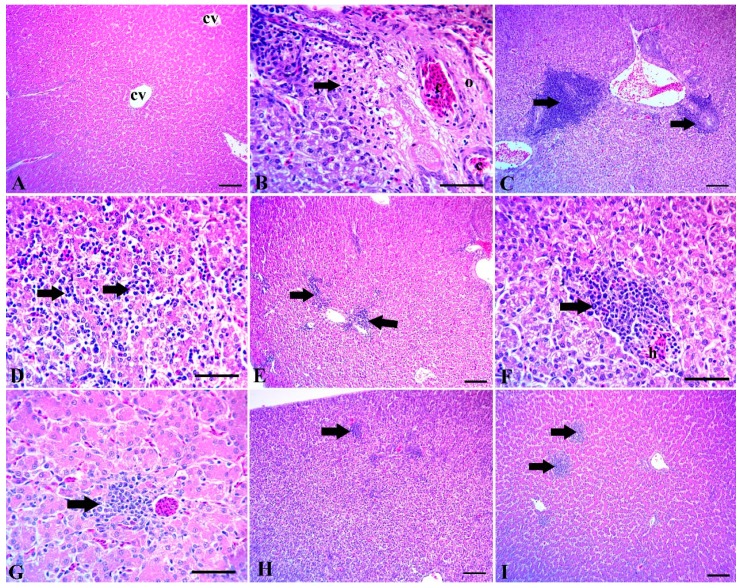
Figure 1.
(A) Liver of control group without Clostridium challenge, showing normal tissue architecture, normal hepatic lobules, a normal central vein (cv) and hepatic sinusoids. (B–D) The intestine of chicks after the Clostridium challenge. (B) Congestion of portal blood vessels (c) with perivascular edema (o) and coagulative necrosis of the surrounding hepatocytes (arrow). (C) Numerous lymphocytic aggregations around the congested portal blood vessels. (D) Extensive lymphocytic aggregations among the hepatocytes (arrows). (E–I) Liver of chicks challenged with Clostridium and supplemented with: (E) Maxus, showing moderate scattered lymphocytic aggregations (arrows); (F) Clostat, showing mild hemorrhage and (H) focal aggregation of lymphocytes (arrow); (G) Sangrovit, showing mild perivascular lymphocytic aggregation (arrow); (H) Clostat + Sangrovit, showing mostly normal hepatic tissue, except for minute focal lymphocytic aggregations (arrow); (I) Gallipro, showing moderate focal lymphocytic aggregations. Scale bar = 100 µm (A,C,E,H,I) and 50 µm (B,D,F,G).

