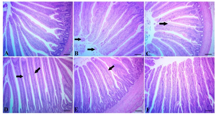
Figure 2.
(A) Intestine of the control group without Clostridium challenge, showing normal tissue architecture. (B) The intestine of chicks supplemented with Maxus showing mild desquamation of villous epithelium (arrow). (C) The intestine of chicks supplemented with Clostat, the columnar epithelium lining the villi into goblet cells (arrow). (D) The intestine of chicks supplemented with Sangrovit, showing moderate metaplasia of the columnar epithelium lining the villi into goblet cells (arrow). (E) The intestine of chicks supplemented with Clostat + Sangrovit, showing mostly normal villi, except for mild metaplasia of the columnar epithelium lining the villi into goblet cells (arrow). (F) The intestine of chicks supplemented with Gallipro, showing normal tissue architecture with normal intestinal villi. Scale bar = 100 µm.

