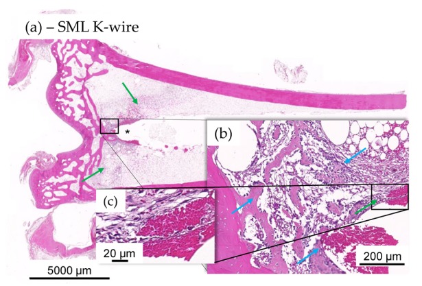
Figure 9.

(a–c) Histology images of distal tibia of test item animal. No evidence of mild ongoing osteomyelitis along the K-wire in most SML K-wire implanted animal and stabilization was often seen to be more significant in the test item at tip of the K-wire imprint (*). Integration was by means of fibroplasia (green arrows) and new fibrous bone formation at host-implant interface ((b), blue arrows), devoid of any heterophilic infiltration (c).
