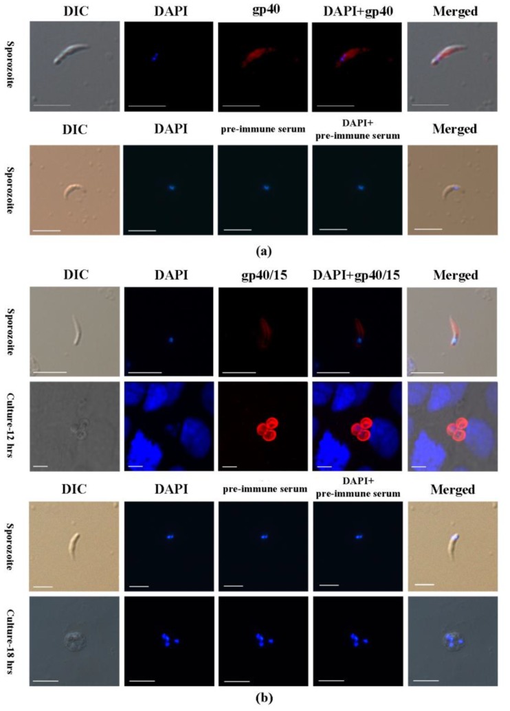
Figure 3.
Immunofluorescence microscopic detection of Cpgp40/15 and Cpgp40 in different C. parvum life cycle stages. (a) Distribution of Cpgp40 in the sporozoites using the rabbit anti-Cpgp40 antibody and pre-immune serum. (b) Association of Cpgp40/15 with the parasitophorous vacuole membrane (PVM) during the parasite intracellular developmental stages, including early and mature meronts, using rabbit anti-Cpgp40/15 antibody and pre-immune serum. Images were taken by differential interference contrast microscopy (DIC), fluorescence microscopy using nuclear stain 4,6-diamidino-2-phenylindole (DAPI); Scale-bars: 5 μm.

