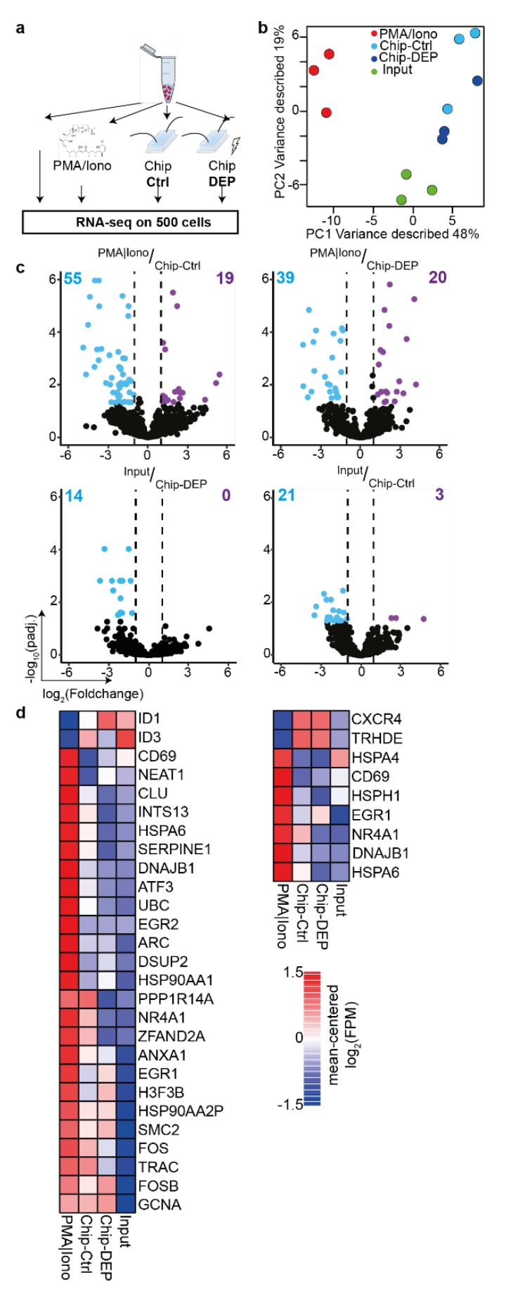
Figure 5.
The electric field has minor molecular impact on Jurkat T-cells. (a) Jurkat cells were either injected into the microfluidics chip (Chip-Ctrl) or additionally subjected to the electric field used for accurate capture and retrieval of cells (Chip-DEP). Controls were either the input cells (Input) or cells activated for three hours under Phorbol-12-myristate 13-acetate and Ionomycin activation (PMA/Iono). Cells from all conditions were cultured for three hours to permit transcriptional changes to take place subsequent to treatment. (b) Principal component analysis on all differentially expressed genes (number of DEGs: 117). (c) Volcano plots of mean RNA-seq FPM (Fragment Per Million) comparing indicated samples. Number of DEGs is indicated. (d) Heatmaps represent expression of selected DEGs. Left: DEGs between PMA/Iono-stimulated and Input cells. Right: DEGs common on comparing Chip-Ctrl and Chip-DEP to PMA/Iono-stimulated cells. Experiments were performed in three independent biological replicates. DEG, differentially expressed gene (absolute(log2[foldchange] >= 1 and padj. <= 0.05).

