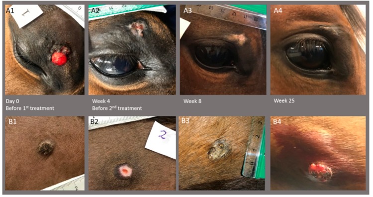
Figure 4.
Images of two sarcoids on horse #3 before the first treatment, before second treatment at week 4, and at follow-up 8 and 25 weeks after first treatment. Top row (A1–4) shows sarcoid placed on the eyelid with complete response (A4; only scar tissue left), and bottom row (B1–4) shows sarcoid placed on the ventral abdomen with the growth of the sarcoid 25 weeks after the first treatment (B4). Note that biopsy of the ventral abdomen sarcoid was performed before images were taken at week 4 (B2).

