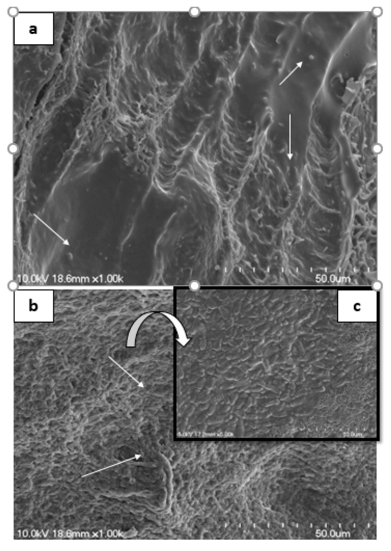
Figure 6.
Scanning electron microscopy (SEM) of excised rat GIT showing presence of large number of coccus-shaped bacteria on the upper surface (G0) (a). The rat administrated with B. longum B-11 displayed huge number of rods on the microvilli of GIT (G1) (b). Magnified images of B. longum B-11 showing bacterial cell are V or Y shaped rods (c).

