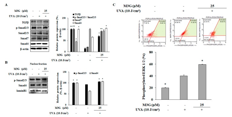
Figure 9.
Effect of M3G on the TGFβ/Smad signaling activation in UVA-irradiated HDFs. Cells were exposed to UVA radiation (10 J/cm2) and treated with 25 μM of M3G. After 24 h incubation cells were harvested and the protein levels of TGFβ, phosphorylated (p-) and inactive Smad 2/3, Smad 7 and Smad 4 in whole cell lysates (A), and p-Smad 2/3 and Smad 4 levels in nuclear fractions (B) were determined using Western blotting. β-actin and lamin B1 (nuclear fraction) was used as an internal control for Western blotting. Activation levels of ERK1/2 in UVA-irradiated HDFs treated and non-treated with M3G (25 μM) were analyzed by flow cytometry and given as percentage of cell population with activated ERK1/2 (C). * p < 0.05, ** p < 0.01 compared to UVA-irradiated untreated control.

