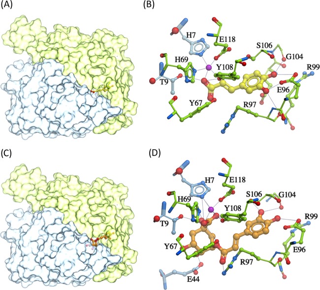
Figure 1.
Docked conformation of caffeic acid/chlorogenic acid. (A) Caffeic acid (yellow sticks) docked within the catalytic site of the dimeric FosX protein; (B) predicted ligand–residue interactions of caffeic acid (yellow) and the FosX protein; (C) chlorogenic acid (orange sticks) docked in the catalytic site of the FosX protein; and (D) molecular interactions of chlorogenic acid (orange) with the FosX protein. Two subunits of the FosX protein are colored distinctly (green and blue); the manganese ion is shown in purple. Hydrogen bonds are represented as black dashes.

