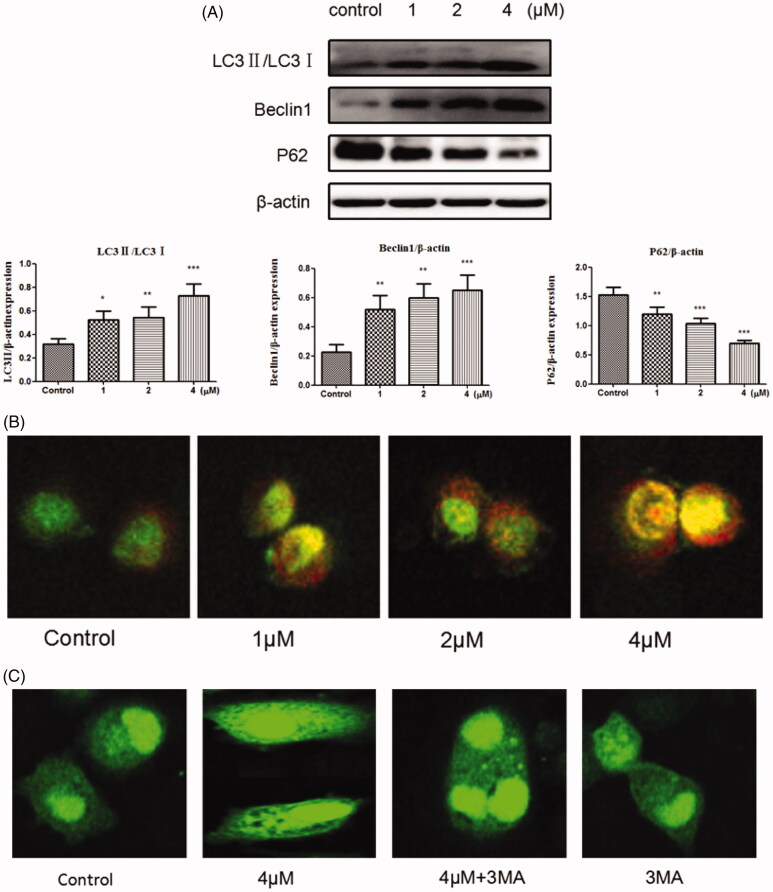
Figure 6.
(A) Western blots assay examined the expressions of LC3-II/LC3-, P62, Beclin1. *p < 0.05, **p < 0.01, ***p < 0.001 compared with the control group. (B) SGC-7901 cells were stained with AO after exposed to compound 12 for 48 h, then detected by the confocal microscopy at 200×. (C) SGC-7901 cells were transfected with GFP-LC3 plasmid, and treated with compound 12 alone (4 μM), 3MA (500 μM, pro-incubated for 1 h), compound 12 and 3MA, then observed under a confocal microscopy at 200×.

