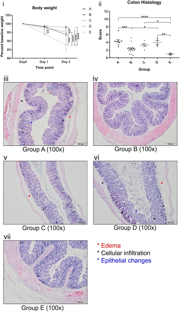
Figure 2.
. (i) Body weight of mice gavaged with different C. difficile ribotypes. (ii) Total histology scores of H&E stained colon tissues of mice at the time of sacrifice (day 2) with different C. difficile ribotypes. Group A, ribotype 002 (n = 8); Group B, other ribotypes, include ribotypes 012, 014, and 046 (n = 21); Group C, ribotype 027 (n = 8); Group D, ribotype 087 (n = 7) of standard strain (VPI10463); Group E, BHI control group (n = 4). *p < 0.05; **p < 0.01; ***p < 0.001; ****p < 0.0001 significantly different between the indicated groups. (iii–vii) Histology photos of H&E stained colon tissues from each group of different ribotypes; Group B and E demonstrate normal colon sections with no significant edema, cellular infiltration and epithelial changes; Group A, C and D show some signs of significant edema, cellular infiltration and epithelial changes when compared to Group B and E.

