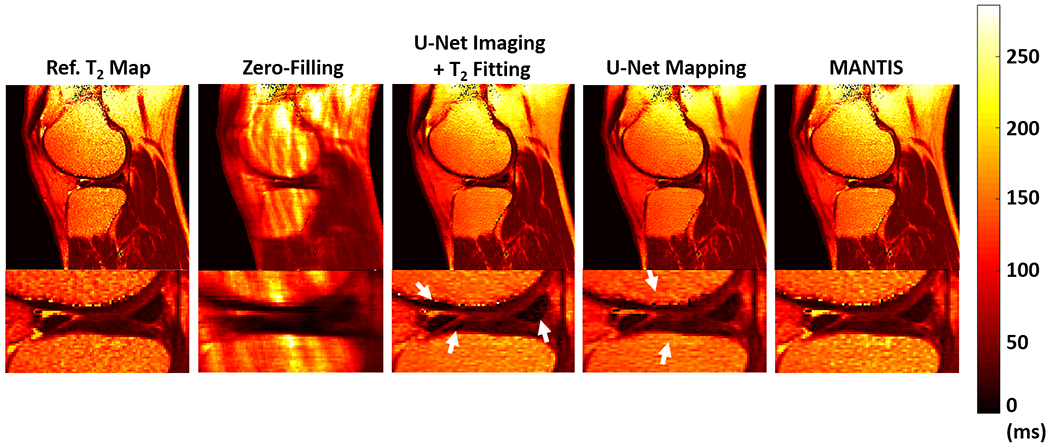
Figure 4:

Comparison of T2 map estimated from MANTIS (direct mapping of the T2, loss1+loss2) with T2 maps generated from U-Net Mapping (direct mapping of the T2, loss2 only) and U-Net Imaging + T2 Fitting approach at R=5. U-Net Mapping is implemented using the MANTIS framework without the loss 1 (data consistency) component. U-Net Imaging + T2 Fitting is a two-step approach in which multiecho images are first generated (loss1 + loss2) followed by parameter fitting. MANTIS achieved better performance (more homogeneous tissue structure and realistic appearance) compared to the other two methods particularly in the cartilage and meniscus where the SNR is typically low.
