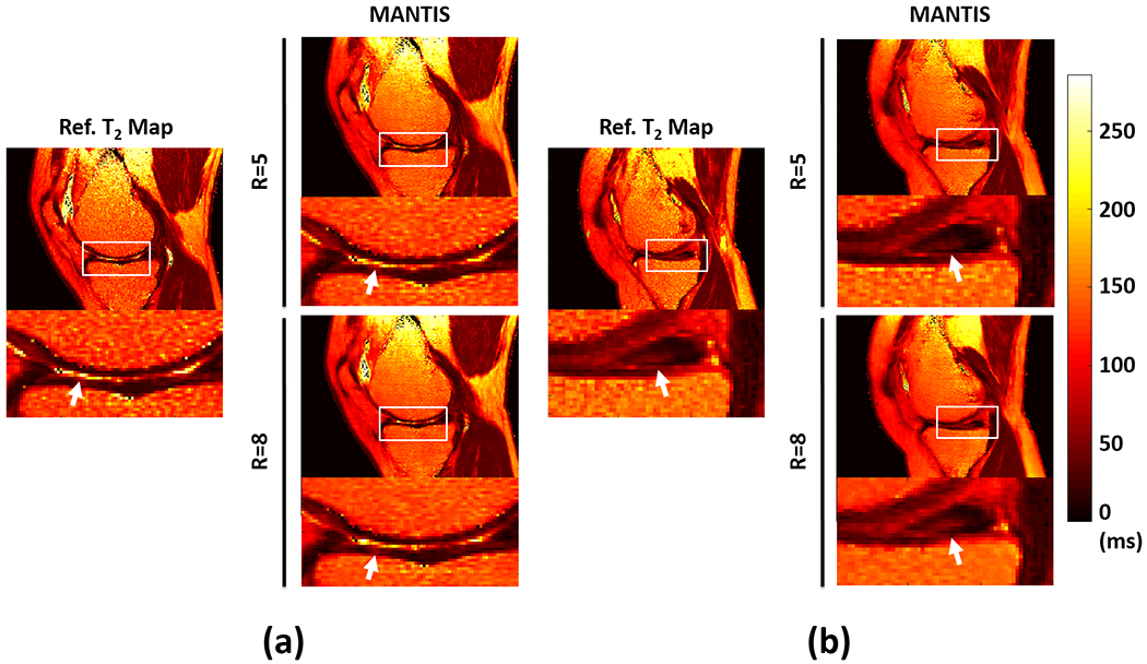
Figure 7:

Two representative examples demonstrating the performance of MANTIS in cartilage and meniscus lesion detection. a) Results from a 67-year male patient with knee osteoarthritis and superficial cartilage degeneration on the medial femoral condyle and medial tibia plateau. b) Results from a 59-year male patient with a tear of the posterior horn of the medial meniscus. MANTIS was able to reconstruct high-quality T2 maps for unambiguous identification of cartilage and meniscus lesions at both R=5 and R=8.
