Figure 1.
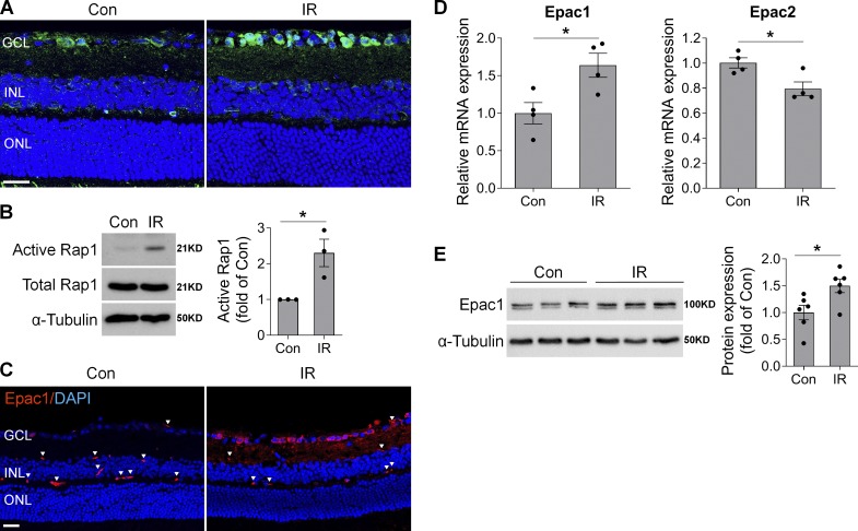
cAMP/Epac1 signaling is increased in mouse model of IR injury. WT mice were subjected to IR injury, and retinas or eyeballs were collected at various times after IR. (A) Representative images of cAMP immunostaining (green) in retinal frozen sections from control (Con) and injured eyes 3 h after IR. Blue, DAPI staining. n = 4 mice. (B) Active Rap1 was assessed by pull-down assay in control and injured retinas 3 h after IR, followed by Western blotting with anti-Rap1 antibody. One representative blot from three independent experiments was shown. Graph represents relative amount of active Rap1. n = 3 mice. (C) Epac1 immunostaining (red) in retinal sections from control and injured eyes 12 h after IR. Blue, DAPI staining. Arrowheads indicate nonspecific staining on vessels. n = 3–4 mice. (D and E) Epac1 and Epac2 mRNA expression by quantitative PCR (n = 4 mice; D) and Epac1 protein expression by Western blotting (n = 6 mice; E) in noninjured control retinas or injured retinas 24 h after IR. *, P < 0.05; Student’s t test. Scale bar: 20 µm. ONL, outer nuclear layer. Error bars represent SEM.

