Figure 10.
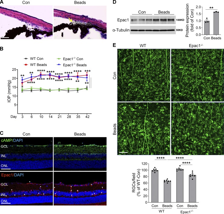
cAMP/Epac1 mediates RGC injury in microbead-induced glaucoma. WT and Epac1−/− mice at 2 mo of age were injected with microbeads into the anterior chamber to induce IOP. (A) Representative images show microbead distribution in the mouse anterior chamber following intracameral injection. (B) IOP elevation in WT and Epac1−/− mice at various time points following microbead injection. n = 8–10; *, P < 0.05; **, P < 0.01; ***, P < 0.001; ****, P < 0.0001; Student’s t test for each time point. (C) The immunoactivity of cAMP (green, upper panel) and Epac1 (red, lower panel) in the retinal sections of WT mice 7 d after microbead injection. Blue, DAPI staining for nuclei. Arrowheads indicate nonspecific staining on vessels. n = 4 mice. (D) Epac1 protein expression in WT retinas 7 d after microbead injection. n = 3; two retinas were pooled for one sample. **, P < 0.01; Student’s t test. (E) Retinal flatmounts from WT and Epac1−/− mice were labeled with Tuj1 antibody (green) 6 wk after microbead injection. n = 7 mice; eight images were taken at the peripheral retina for each sample and calculated as average value; ****, P < 0.0001; one-way ANOVA. Scale bar: 50 µm. ONL, outer nuclear layer. Error bars represent SEM.

