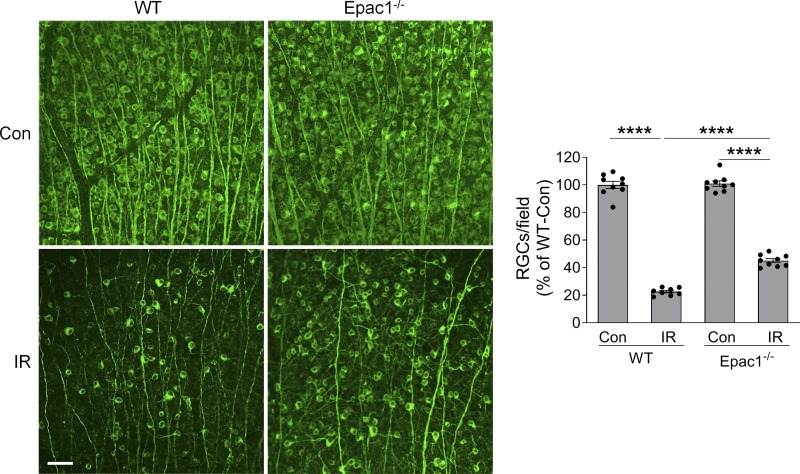
Figure S3.
Epac1 deletion prevents RGC loss 14 d after IR. Representative images of retinal flatmounts labeled with Tuj1 antibody (green) 14 d after IR in WT and Epac1−/− mice. Bar graph represents the number of Tuj1-positive cells per field. Scale bar: 50 µm. n = 8–9 mice; eight images were taken at the peripheral retina for each sample and calculated as average value. ****, P < 0.0001; one-way ANOVA. Error bars represent SEM.

