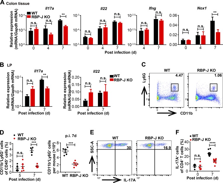
Figure 2.
RBP-J KO mice display impaired colonic Th17 cell immune responses upon C. rodentium infection. WT and RBP-J KO mice were orally inoculated with 2 × 10⁹ CFUs of C. rodentium, and tissues and LP were harvested at the indicated time points p.i. (A and B) qPCR analysis of the indicated mRNAs in colon tissues (A) and LP mononuclear cells (B). (C and D) Colonic LP CD11b+Ly6G+ neutrophils were determined by flow cytometry analyses (FACS). Representative FACS plots at day 7 p.i. (C) and cumulative data of cell ratio (D, [left]) and absolute numbers (D, [right]) at the indicated p.i. days are shown. (E and F) Colonic LP mononuclear cells at day 7 p.i. were treated with PMA and ionomycin in vitro for 4–5 h, and representative FACS plots of IL-17A production in LP CD3+CD4+ T cells are shown. Data are pooled from two independent experiments (A, B, D, and F); n ≥ 3 in each group. Data are shown as mean ± SEM; n.s., not significant; **, P < 0.01; ***, P < 0.001; ****, P < 0.0001; two-tailed Student’s unpaired t test. Each symbol in D and F represents an individual mouse. SSC-A, side scatter area.

