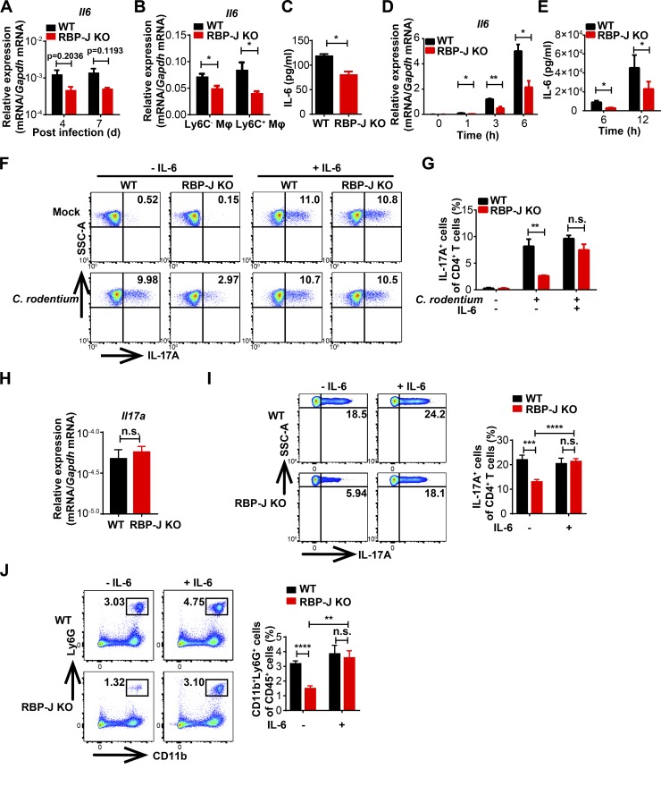
Figure 3.
Macrophage-intrinsic RBP-J promotes Th17 cell immune responses via sustaining IL-6 expression. (A–C) WT and RBP-J KO mice were orally inoculated with 2 × 10⁹ CFUs of C. rodentium. Colonic LP mononuclear cells were isolated at day 5 p.i. Data are pooled from two independent experiments; n = 3 in each group. (A) qPCR analysis of Il6 in colon tissues at the indicated p.i. days. (B) qPCR analysis of Il6 in sorted CD64+Ly6C− and CD64+Ly6C+ colonic macrophages. (C) IL-6 levels in the supernatants of sorted CD64+Ly6C− colonic macrophages (2 × 10⁶ cells per ml) were measured by ELISA. (D and E) IL-6 levels of peritoneal macrophages stimulated with heat-killed C. rodentium (MOI = 1) were determined by qPCR (D) and ELISA (E). Data are pooled from three independent experiments. (F and G) Naive CD4+ T cells were sorted and cultured with the supernatants of peritoneal macrophages that were not treated (mock) or stimulated with heat-killed C. rodentium (MOI = 1) and TGF-β, without (left) or with (right) IL-6 for 72 h. FACS (F) and cumulative data (G) of IL-17A–expressing CD4+ T cells are shown. Data are pooled from three independent experiments. (H–J) 6–8-wk-old WT and RBP-J KO mice were intraperitoneally injected with recombinant IL-6 at days 3 and 5 p.i. and sacrificed at day 7 p.i. Data are pooled from two independent experiments, n ≥ 4 in each group. (H) qPCR analysis of Il17a in colon tissues. (I and J) Representative FACS plots (left) and cumulative data (right) of IL-17A production in LP CD3+CD4+ T cells (I) and colonic LP neutrophils (J). Data are shown as mean ± SEM; n.s., not significant; *, P < 0.05; **, P < 0.01; ***, P < 0.001; ****, P < 0.0001; two-tailed Student’s paired t test (D and E) or Student’s unpaired t test (other panels).

