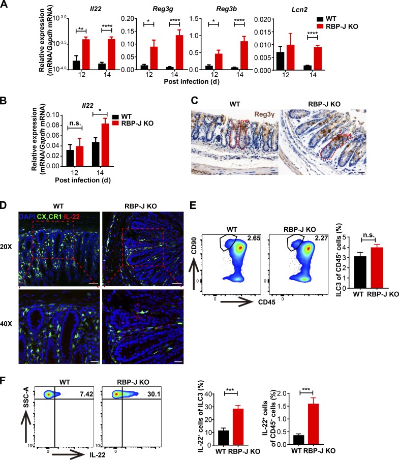
Figure 6.
RBP-J KO mice display heightened levels of intestinal ILC3-derived IL-22 during the late phase of infection. WT and RBP-J KO mice were orally inoculated with 2 × 10⁹ CFUs of C. rodentium. (A and B) qPCR of Il22 and AMP mRNAs in colon tissues (A) and LP mononuclear cells (B). Data are pooled from two or three independent experiments; n ≥ 3 in each group. (C) Immunohistochemical analysis of Reg3γ protein levels at day 14 p.i. (scale bars represent 20 µm). (D) Immunofluorescence staining for IL-22 (red) and DAPI (blue) in the distal colon from WT (CX3CR1gfp/+ Lyz2-Cre) and RBP-J KO (CX3CR1gfp/+ Rbpjfl/fl Lyz2-Cre) mice at day 14 p.i. Scale bars represent 50 µm (top panels) and 20 µm (bottom panels). (E and F) Representative FACS plots (left) and cumulative data (right) of colonic LP ILC3 (CD45midCD3−Thy-1+) populations (E) and IL-22 production in ILC3 (F) at day 14 p.i. Data are pooled from three independent experiments; n ≥ 3 in each group. Data are shown as mean ± SEM; n.s., not significant; *, P < 0.05; **, P < 0.01; ***, P < 0.001; ****, P < 0.0001; two-tailed Student’s unpaired t test. SSC-A, side scatter area.

