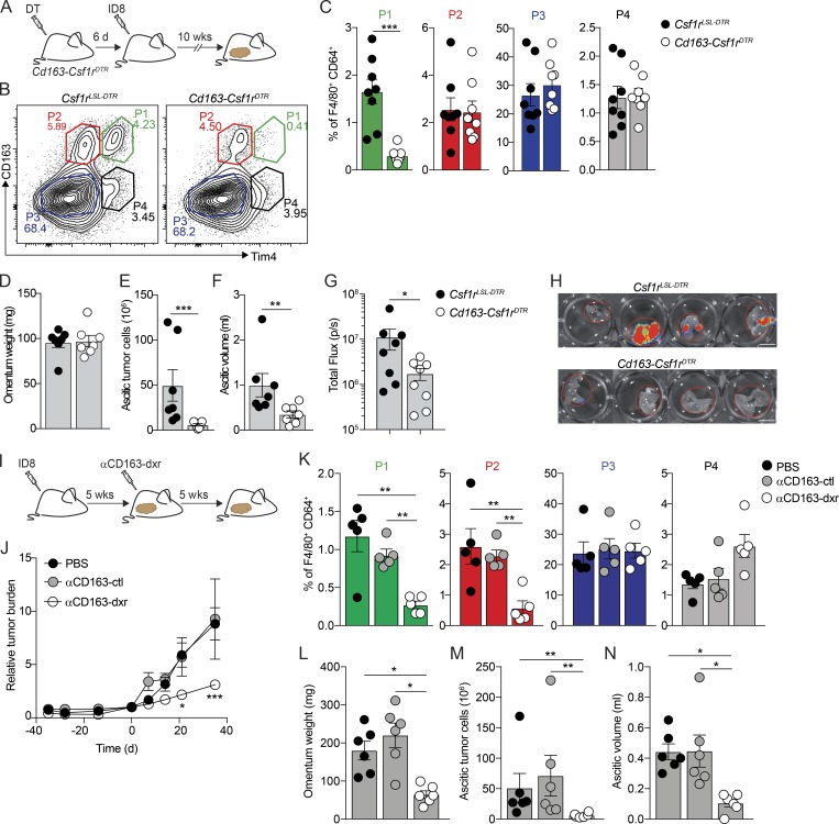
Figure 6.
Specific depletion of CD163+ Tim4+ tissue-resident macrophages prevents metastatic spread of ovarian cancer. (A) Cohorts of Cd163-Csf1rDTR or control mice (Csf1rLSL-DTR) were injected with 4 ng/kg DT 6 d before transplantation with ID8 cells. Mice were analyzed for depletion of macrophages and effects on tumor growth at 10 wk. (B and C) Flow cytometry analysis of CD163hi Lyve-1+ macrophages in omentum of Cd163-Csf1rDTR and Csf1rLSL-DTR mice 10 wk after injection of ID8 cells. (D–F) Omentum weight (D), total tumor cells in ascites (E), and ascites volume (F) in Cd163-Csf1rDTR and Csf1rLSL-DTR mice treated with DT. (G and H) Ex vivo bioluminescence analysis of metastases on the diaphragm of Cd163-Csf1rDTR and Csf1rLSL-DTR; scale bar: 0.5 cm. Data are represented as mean ± SEM of n = 7, and statistically significant difference was calculated using Mann-Whitney U test; *, P < 0.05; **, P < 0.01; ***, P < 0.001. (I) Therapeutic depletion of CD163+ macrophages; mice were injected with ID8 cells and after 5 wk randomized into groups and treated with dxr-loaded αCD163-LNPs (αCD163-dxr), empty αCD163-LNPs (αCD163-ctrl), or PBS alone twice a week for 5 wk. (J) Tumor burden monitored by in vivo bioluminescent imaging. Data are represented as mean ± SEM of n = 6, and statistically significant difference was calculated using two-way ANOVA followed by Tukey post hoc test; *, P < 0.05; ***, P < 0.001. (K) Flow cytometry analysis of P1–P4 macrophages in omentum after therapeutic depletion of CD163+ cells. (L–N) Omentum weight (L), total tumor cells in ascites (M), and ascites volume (N). Data are represented as mean ± SEM of n = 6, and statistically significant difference was calculated using Kruskal-Wallis one-way ANOVA followed by Dunn’s multiple comparisons test; *, P < 0.05; **, P < 0.01; ***, P < 0.001. All data are representative of three independent experiments.

