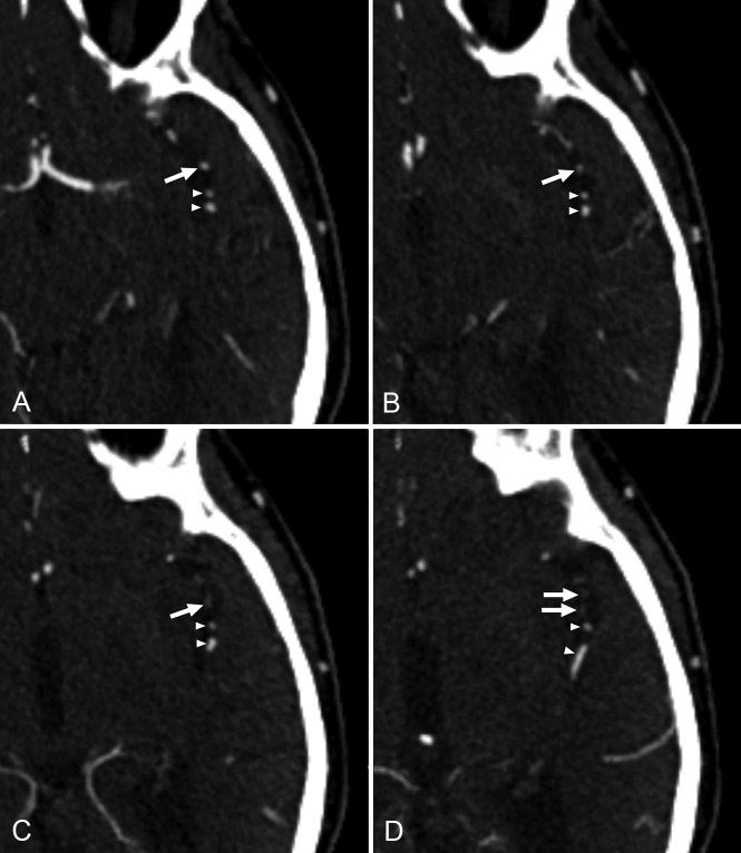
Fig 1.
A 75-year-old male patient with acute ischemic stroke. At initial CTA evaluation, occlusion of 1 of the M2 segment branches of the left middle cerebral artery (arrows on all slices) was missed. Consecutive axial CTA slices in a caudocranial direction (A–D) show a contrast filling defect in a branch of the left M2 segment (arrows in C and D). Note that 2 adjacent branches of the left M2 segment show normal contrast filling on all slices (arrowheads).

