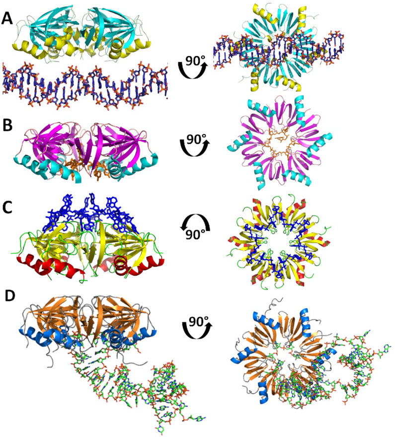
Figure 2.

Hfq has three distinct nucleic acid binding sites. (A) Side view of the Hfq–DNA complex. Ninety-degree rotation shows view looking into the proximal face. (B) Side view of AU5G RNA (orange) shown bound in the proximal pore of Staphylococcus aureus Hfq. Ninety-degree rotation shows view into the proximal face. (C) Side view of A15 RNA (blue) shown bound to the distal face of Escherichia coli Hfq. Ninety-degree rotation shows view into the distal face. (D) Side view of RydC RNA (green) bound to the proximal face of E. coli Hfq. Ninety-degree rotation shows view into the proximal face.
