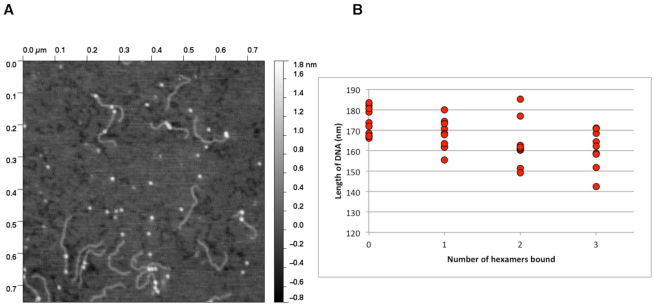
Figure 5.
The binding of multiple Hfq(2-69) hexamers compacts dsDNA. (A) A representative AFM topograph demonstrating a 500 bp DNA (the Escherichia coli hipBA promoter region) bound by one or more Hfq hexamers (bright spots). (B) Representative scatter plot of DNA measurements made in the presence and absence of Hfq from a series of AFM topographs. Each point is the average of three measurements of the length of DNA molecules where the number of Hfq hexamers bound was obvious, i.e. Hfq was not bound at the ends or multiple hexamers were clustered together preventing delineation of the number bound to the DNA.

