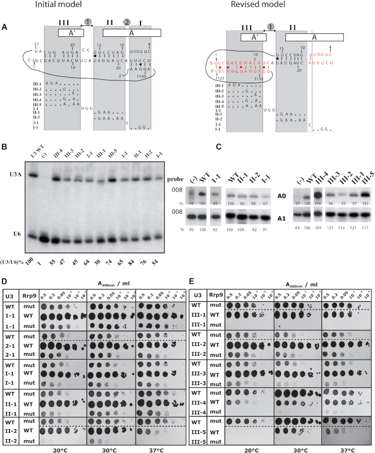
Figure 8.
The Rrp9-R289A mutation is synthetic lethal with some of the U3 snoRNA mutations destabilizing the formation of the helices formed between U3 and the 18S pre-rRNA (10,17,28,30). (A) Representation of the initial and new models for U3 snoRNA/pre-rRNA base-pair interactions. In the initial model, the three helices (I, II, III) and segments (1,2) are represented. In the new model, helix II which is unchanged and the new version of helix III (in red) are shown, as well as the opened helix I (in red) (40,44). The mutations tested in the synthetic lethality screen are indicated below each model. For the complete set of mutations, see Supplementary Table S3. (B) Northern-blot analysis of the in vivo stability of the different U3 mutants indicated on top of the autoradiogram. They were expressed in JH84 yeast cells grown at 30°C in YPD medium. About 20 μg of total RNA were fractionated by electrophoresis on a 6% denaturing polyacrylamide gel. After electrotransfer on a nylon membrane, the U3 snoRNA variants were detected by hybridization of the membrane with the 5′-end labeled U3 probe and for internal control, with the U6 oligonucleotide, complementary to U6 snRNA (Supplementary Table S1). For each U3 variant, the ratio between the U3 and U6 signals of the autoradiogram were calculated and expressed as a percentage of the value obtained for WT U3 arbitrary taken as 100%. (C) Primer extension analysis of total RNA extracted from JH84 cells expressing WT or variant U3 snoRNA, grown on YPD medium, The labeled 008 primer, complementary to 18S rRNA (Figure 3A) was used as the probe. The signals corresponding to the A0- and A1-ending products are shown and their relative amounts were expressed as percentages of the amount obtained for WT U3 arbitrary taken as 100%. (D, E) Yeast cells were transformed with plasmids expressing wild-type Rrp9 (WT) or R289A Rrp9 mutant (mut) and U3 snoRNA (Wild-type WT or mutants 1-1, 2-1, I-1, II-1, II-2, III-1 to III-5) and grown on YPD medium after serial dilutions (2, 5, 20, 102, 103 or 104 X) at 20, 30 and 37°C (left, middle and right panels, respectively; for details see methods section). A comparative analysis of cell growth on YPD and YPG media is shown in the Supplementary Figure S5.

