Figure 5.
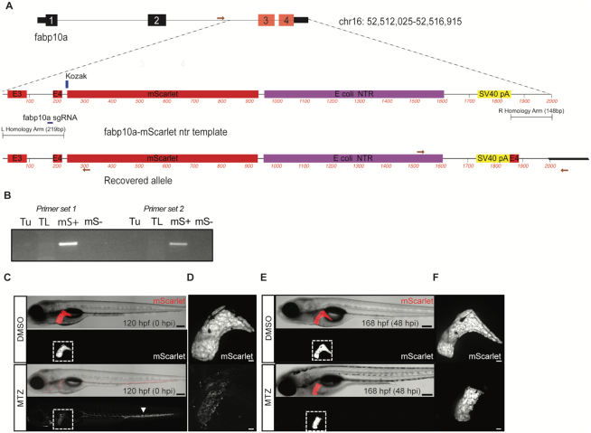
Knock-in of a synthetic dsDNA template encoding a fluorophore and bacterial nitroreductase (ntr) enables tissue-specific cellular ablation. (A) Schematic depicting fabp10a locus and fabp10a-mScarlet ntr template for HDR. A schematic of the recovered allele from F1 progeny confirms integration of mScarlet after the last coding exon of fabp10a and includes a duplicated exon 4. Primers used for PCR are displayed as red arrows. (B) Allele-specific PCR was performed to confirm integration of the template in mScarlet+ (mS+) embryos. (C–F) Mtz exposure induced hepatocyte injury in fabp10a-mScarlet-NTR larvae. (C) Confocal imaging revealed loss of fluorescence in the liver (outlined) of Mtz-treated but not DMSO-treated larvae. Remnants of ablated hepatocytes were found distributed throughout the vasculature (arrowhead). (D) Confocal imaging of liver in DMSO- and Mtz-treated larvae after injury. (E) At 48 h post injury (hpi), mScarlet signal was observed in the liver (outlined) of the Mtz-treated larvae, demonstrating liver regeneration. (F) Confocal imaging of liver in DMSO and Mtz treated larvae at 48 hpi. Scale bars are 200 μm (C, E) and 30 μm (D, F).

