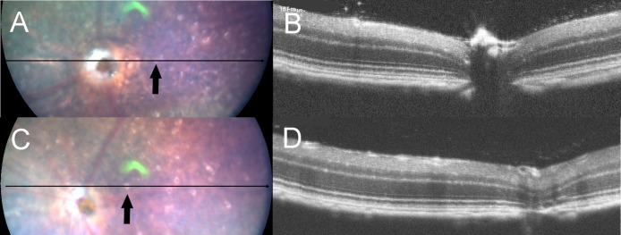
Fig 3. The fundus photographs (panels A and C) and corresponding OCT images (panels B and D) of Rdh5-/- mice.
Panels A and B were taken at PM4, and panels C and D were recorded at PM6. The lines in the left panels A and C indicate the section line of the corresponding OCT images in the right panels B and D, respectively. Note that the orientations of B and D are opposite to those of A and C. Arrows indicate the location of white spots.

