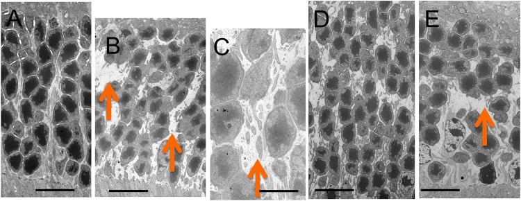
Fig 7. Electron microscopic findings of the outer nuclear layer of C57BL/6J (A) and Rdh5-/- (B and C, respectively) mice at PM4, and C57BL/6J (D) and Rdh5-/- (E) mice at PM6, respectively.
Arrows indicate wide interspaces between nuclei of the photoreceptors. Bars indicate 10 μm in panels A, B, D and E, and 2 μm in panel C.

