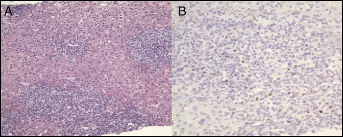
Figure 2.
Histopathologic images of the patient's acute liver injury from liver biopsy showing (A) there is severe acute hepatitis, consistent with an Epstein-Barr virus-driven hepatitis. There is a marked mixed portal and lobular inflammatory infiltrate showing numerous inflammatory cells in the hepatic sinusoids. Marked cholestasis is present, and apoptotic hepatocytes are present (hematoxylin & eosin stain 200×). (B) Highlighted scattered nuclear staining in the lymphocytes, which is compatible with Epstein-Barr virus infection (hematoxylin & eosin stain 400×).

