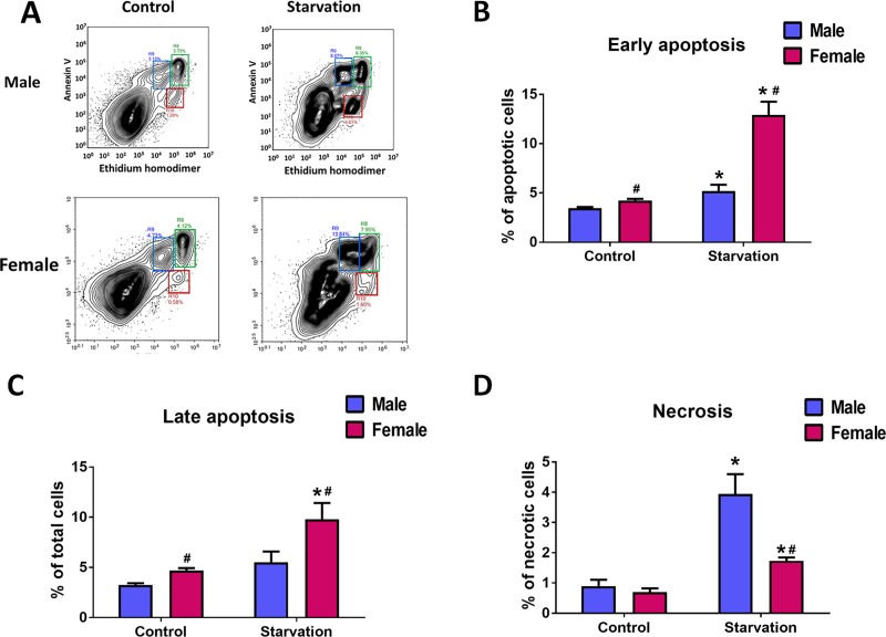
Fig 6. Sex-specific profile of apoptotic and necrotic cell death in HPAEC.
Stress in human endothelial cells obtained from age and race matched male and female donors was induced by culturing HPAEC in serum-free media for 48h. A—representative plots showing populations of Annexin V and EthD-1 positive cells in Control and starved male and female HPAEC. B-D–quantitative analysis of early apoptosis, late apoptosis, and necrosis in male and female HPAEC. Values are means ± SEM. N = 6 for each sex. *p < 0.05 versus same-sex Control cells; #p < 0.05 versus male HPAEC. Statistical analysis was performed using unpaired t-test.

