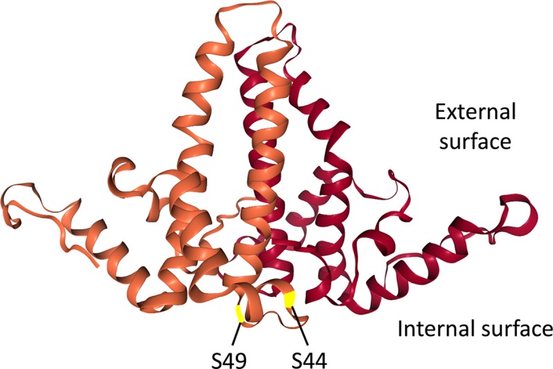
Fig 1. HBc dimer structure and location of the putative NTD phosphorylation sites S44 and S49.

The structure of the two HBc NTD monomer (in brown and dark red, respectively) in an NTD dimer, based on the HBV capsid crystal structure [17], is shown. The two putative NTD phosphorylation sites S44 and S49, located on the interior surface of the capsid, are highlighted.
