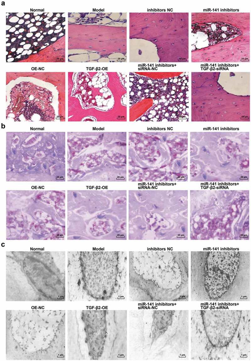
Figure 3.

Histopathological observation of femoral head tissues in ONFH rats in each group. (a) HE staining of femoral head tissues of rats in each group; (b) Masson staining of femoral head tissues of rats in each group; (c) Electron microscopic ultrastructure of femoral head tissues of rats in each group.
